Secret Life of the Unborn Shark
Part two
How to Build a Shark a Closer Look
|
Let's take a closer look at the developmental events leading up to this stage. Recall from our Overview that the blastodisc rolls up on itself. This process is termed folding of the neural fold. Strange as it might seem, the spinal chord and its large anterior expansion the brain start out on the outside of the embryo. Folding the neural fold allows creation of a hollow tube of nervous tissue. This may be particularly important to create a series of sinuses (cavities) through which a special fluid (cerebrospinal fluid) can circulate in the brain, so that this relatively large and complex mass of nervous tissue can regulate itself hormonally.
|
|
Here's a diagramatic view of the developing blastodisc as seen from the side, showing how a flat plate of undifferentiated animal cells begin to fold themselves into a shark. The colors indicate which parts of the blastodisc differentiate into various tissues. The yellow part develops into the notochord, which is a stiffening cartilaginous rod that forms the scaffolding for the backbone, which eventually will be differentiated into segmental vertebrae. The red develops into the muscle blocks, those of which along the flanks will develop into swimming muscles. The green part become the gut, consisting of esophagus, stomach, intestine (which, in sharks, is internally partitioned), rectal gland (a salt-secreting organ unique to elasmobranchs), and rectum. The blue part develops into the brain, which coordinates nervous activity and is quite large in sharks. As the blastodisc develops, it rolls under itself like the caterpillar tracks of a tank, eventually meeting up with the head end to form a three-layered embryo.
Here is a diagramatic side-view of the anterior of a developing dogfish embryo. At a later stage of development, the embryo continues to differentiate and starts taking on an increasingly shark-like form. Note that the nerve tube (purple) and eye (grey) has started to differentiate from the brain. Note also that the coelom, or body cavity (orange), has started to form.
More details are visible in this three-dimensional cutaway diagram. Here, you can see the nerve tube (purple) is indeed hollow and the notochord (yellow) below it. You can also see the muscle blocks (red) start to differentiate and its spatial relationship to the coelom.
Here we have a diagrammatic look at a developing dogfish embryo, showing all the parts we saw in the earlier photographs.
Closing Thoughts
 All of the foregoing is biologically fascinating. In truth, there are many
more embryological details into which I could have gone but on which I did not
even touch. But I'd like to close on a more philosophical note.
All of the foregoing is biologically fascinating. In truth, there are many
more embryological details into which I could have gone but on which I did not
even touch. But I'd like to close on a more philosophical note.
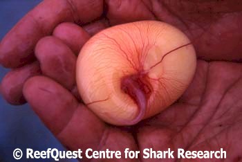
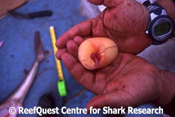
As a naturalist and a lover of sharks, it was very sad for me to see the dead mother Green-Eyed Dogfish and her seven unborn embryos. With one capture, eight lives were cut short. It is a strange feeling to hold an unborn shark in the palm of your hand to realize that what you are holding had the potential to develop into a sleek, agile marine predator that might have gone on to create more young, had we not come along and ended its mother's life. But there is a very big difference between potential and actual. The seven embryos were not really sharks ... not yet, at any rate. They were merely potential sharks. But the mother dogfish was an actual shark, going about her own business until she had the misfortune to take our captain's hook.
It would be a mistake to think ill of the man who killed them all. The vessel captain who caught the mother dogfish is a very decent fellow who would never kick a dog or strangle a bunny. He is a commercial lobster fisherman, a tough, no-nonsense man who loves his family and works hard to earn a living. He is smart and well read and has a wonderful sense of humor. But, to him, the mother dogfish was just a fish: something to be caught for food or sale. He didn't know she was pregnant and I don't know if that would have made a difference as to whether he let it go. I suspect he retained it because I was on board and he knew I was a biologist interested in sharks. Otherwise, he probably would have cut it up to use as bait for tastier or more saleable fish.
The mother dogfish had in her stomach dozens of lenses from the eyes of teleost fishes and part of a crustacean carapace. She was a professional killer. There is nothing inherently 'evil' or 'immoral' in this way of life. It is just what dogfish do. And catching fish is simply what fishermen do. Sharks gotta eat and so do fishermen. When a fisherman encounters a shark on his terms, the shark invariably loses. It is strange that when a shark catches a human in its environment and bites or kills him, we cannot forgive the creature for simply doing what it does.
 I thought about such things as I held the last of the lifeless Green-Eyed
Dogfish embryos in my hand. I suppose I could have been outraged that the
mother shark and all her young were killed and refused to have anything further
to do with them. But I am a biologist. Studying organisms is what I
do. And besides, not studying that dogfish specimen would have done
nothing to bring it or its young back to life. So I did my best to collect
as much information as I could about the mother shark and her embryos.
Doing so and sharing much of that information here is my way of 'honoring' the
life of the mother dogfish and her unborn young. I hope that this does not
seem unduly sentimental or 'strange'. But mostly, I hope you will have
found these pages interesting and useful.
I thought about such things as I held the last of the lifeless Green-Eyed
Dogfish embryos in my hand. I suppose I could have been outraged that the
mother shark and all her young were killed and refused to have anything further
to do with them. But I am a biologist. Studying organisms is what I
do. And besides, not studying that dogfish specimen would have done
nothing to bring it or its young back to life. So I did my best to collect
as much information as I could about the mother shark and her embryos.
Doing so and sharing much of that information here is my way of 'honoring' the
life of the mother dogfish and her unborn young. I hope that this does not
seem unduly sentimental or 'strange'. But mostly, I hope you will have
found these pages interesting and useful.
