Secret Life of the Unborn Shark
On a recent expedition to the Chatham Islands, New Zealand, I had opportunity to study the early development of spiny dogfishes. An individual dogfish was caught by our vessel's Captain just after midnight on 6 November 2003, while I and most of our crew slept on-board at our overnight anchor site near an offshore rocky islet named Star Keys. It was caught on a handline baited with Blue Moki (Latridopsis ciliaris) over a sandy-rocky bottom about 12 metres deep.
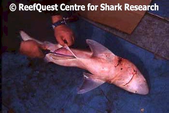
I learned about the catch at first light. I admit that I was rather saddened that this innocent creature had been killed for sport, but the biologist in me was grateful that the carcass was retained and still quite fresh. When I examined the specimen, I determined it was a female Green-Eyed Dogfish (Squalus mitsukurii) measuring 95.6 cm in length. Her abdomen was not notably swollen and thus she was not obviously pregnant. However, from what I had read about this species, I suspected that she was sexually mature.
 |
 |
After taking a series of standard measurements, termed "morphometrics", of her external features, I prepared to dissect her so that I could verify her maturity state, examine her stomach contents, and check for internal parasites. I began by carefully slicing open the belly, from cloaca to between her pectoral fins.
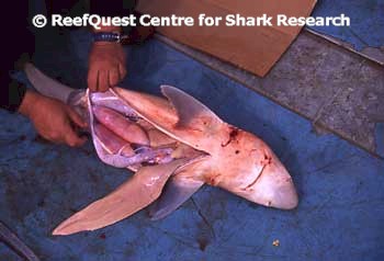
In Green-Eyed Dogfishes, the liver is an enormous organ that might account for 25% of the shark's total weight. I gently pulled out the left lobe of her liver, revealing her reproductive tract. It was immediately apparent that she was pregnant. Low in the belly, her paired uteri were swollen with numerous ripe eggs, each about the size of a small billiard ball. Higher in her abdomen deep in the chest were the ovaries, their surface rough with large, well-formed ova of various sizes.
Overview of Dogfish Development
 All
modern sharks practice internal fertilization. Like other spiny dogfishes
(family Squalidae), female Green-Eyed Dogfish produce large, yolky eggs
called ova. These ova erupt from the ovarian surface when
they are ripe, travel tailward down the Mullerian ducts until they reach
a swelling called the nidamental gland. In the nidamental gland,
the eggs are fertilized by sperm stored from an earlier copulation and then
packaged in a membranous envelope called a candle. The candles are
transferred further rearward to the uteri, where development continues
for 12 to 24 months (depending on water temperature) until the fully-formed
pups are ready to be born alive.
All
modern sharks practice internal fertilization. Like other spiny dogfishes
(family Squalidae), female Green-Eyed Dogfish produce large, yolky eggs
called ova. These ova erupt from the ovarian surface when
they are ripe, travel tailward down the Mullerian ducts until they reach
a swelling called the nidamental gland. In the nidamental gland,
the eggs are fertilized by sperm stored from an earlier copulation and then
packaged in a membranous envelope called a candle. The candles are
transferred further rearward to the uteri, where development continues
for 12 to 24 months (depending on water temperature) until the fully-formed
pups are ready to be born alive.
 My
dissection of this pregnant Green-Eyed Dogfish provided a fascinating 'snapshot'
of one stage of this entire process. A number of important developmental
events occurred before our specimen was caught and development of her young
interrupted. At the site of fertilization there develops a plate of cells
termed a blastodisc (A),
which will eventually develop into
an individual dogfish. The blastodisc sort of folds up on itself (B), from
back to front, to form what will develop into the cartilaginous backbone and
associated structures. Continuing to develop from tail to head (C), the
backbone differentiates into a skull at
the front and adds an arch above that protects the spinal chord
and another arch below that protects the body's main blood vessel, the
dorsal aorta. As all this is going on, the
embryo develops a stalk separating it from
the yolk sac and a network of blood
vessels to transport yolk that fuels its
development (D). As the embryo develops, it continues to add complexity
and becomes a fetus, using up its yolk
supply as it grows (E). Our dissection caught this process at roughly
stage D.
My
dissection of this pregnant Green-Eyed Dogfish provided a fascinating 'snapshot'
of one stage of this entire process. A number of important developmental
events occurred before our specimen was caught and development of her young
interrupted. At the site of fertilization there develops a plate of cells
termed a blastodisc (A),
which will eventually develop into
an individual dogfish. The blastodisc sort of folds up on itself (B), from
back to front, to form what will develop into the cartilaginous backbone and
associated structures. Continuing to develop from tail to head (C), the
backbone differentiates into a skull at
the front and adds an arch above that protects the spinal chord
and another arch below that protects the body's main blood vessel, the
dorsal aorta. As all this is going on, the
embryo develops a stalk separating it from
the yolk sac and a network of blood
vessels to transport yolk that fuels its
development (D). As the embryo develops, it continues to add complexity
and becomes a fetus, using up its yolk
supply as it grows (E). Our dissection caught this process at roughly
stage D.
The Female Dogfish's Reproductive Organs
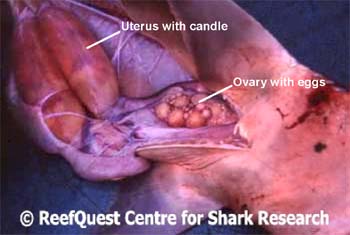
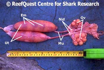 This photo shows the entire reproductive tract of the mature
female Green-Eyed Dogfish I dissected in the Chatham Islands. For
orientation, the anterior (head) end is at the right. Note the paired
ovaries (Ov), with large, well differentiated ova (the largest are
measured 1.8 cm in diameter) and smaller developing ova (each about the size
of a tomato seed) on the ovarian surface. Mullerian ducts (Mul)
transport eggs from the ovaries to the paired uteri (Ut), the two
large, sausage-shaped organs. The swelling in each Mullerian duct just
tailward of the ovaries are the nidamental glands (Nid), where
fertilization takes place. The uterine wall is translucent, so that
within each you can just make out the developing embryos (Em) and
transverse partitions marking the edges of each individual embryo's yolk
supply. The right uterus contains four embryos, the left one only three.
This photo shows the entire reproductive tract of the mature
female Green-Eyed Dogfish I dissected in the Chatham Islands. For
orientation, the anterior (head) end is at the right. Note the paired
ovaries (Ov), with large, well differentiated ova (the largest are
measured 1.8 cm in diameter) and smaller developing ova (each about the size
of a tomato seed) on the ovarian surface. Mullerian ducts (Mul)
transport eggs from the ovaries to the paired uteri (Ut), the two
large, sausage-shaped organs. The swelling in each Mullerian duct just
tailward of the ovaries are the nidamental glands (Nid), where
fertilization takes place. The uterine wall is translucent, so that
within each you can just make out the developing embryos (Em) and
transverse partitions marking the edges of each individual embryo's yolk
supply. The right uterus contains four embryos, the left one only three.
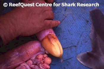 Here I am carefully removing the candle from the right
uterus. This is very delicate work, because the individual eggs
are bound by a very thin, transparent membrane, that feels turgid like a
water balloon filled to near-bursting. Just a tiny bit too much force
and the eggs would rupture, resulting in an undifferentiated yolky mess and
loss of the opportunity to examine the embryos further.
Here I am carefully removing the candle from the right
uterus. This is very delicate work, because the individual eggs
are bound by a very thin, transparent membrane, that feels turgid like a
water balloon filled to near-bursting. Just a tiny bit too much force
and the eggs would rupture, resulting in an undifferentiated yolky mess and
loss of the opportunity to examine the embryos further.
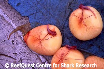
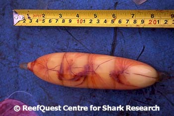 Success! Here we see the entire candle from the
right uterus. You can clearly see the four embryos, their network of
blood vessels, and their yolk supply through the transparent membrane.
Each embryo is separated from its neighbors by transverse membranes.
Let's take a closer look ...
Success! Here we see the entire candle from the
right uterus. You can clearly see the four embryos, their network of
blood vessels, and their yolk supply through the transparent membrane.
Each embryo is separated from its neighbors by transverse membranes.
Let's take a closer look ...
The Developing Embryos
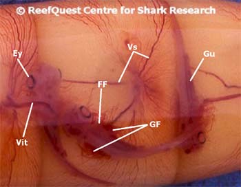 Close up of developing Green-Eyed Dogfish embryos,
photographed through the transparent membrane of the candle. Note that
the eyes (Ey) are large and well developed, with a dark ring around
each pupil. You can also see the dark tube that will develop into the
gut (Gu), the dark blood vessels (Vs) spider-webbing over the
surface of each yolk and extending up the vitelline chord (Vit) to
the developing embryo. (The vitelline chord is analogous to the
umbilical chord in mammals, but it is composed of different tissues and, in
dogfishes, does not interdigitate with the uterine lining to assist in the
uptake of nutrition supplied by the mother; in dogfishes, the entire
development of each pup is fuelled by the yolk supply). Note also the
external gill filaments (GF), which extend outside the gill slits and
into the vitelline fluid to take up dissolved oxygen and dump carbon
dioxide. Lastly, note that the fins are not fully differentiated,
consisting of simple fin folds (FF) without any hint of claspers that
might enable me to determine their sex. The sum-total of these
developmental hallmarks allow me to determine that these embryos are about
5.5 months old.
Close up of developing Green-Eyed Dogfish embryos,
photographed through the transparent membrane of the candle. Note that
the eyes (Ey) are large and well developed, with a dark ring around
each pupil. You can also see the dark tube that will develop into the
gut (Gu), the dark blood vessels (Vs) spider-webbing over the
surface of each yolk and extending up the vitelline chord (Vit) to
the developing embryo. (The vitelline chord is analogous to the
umbilical chord in mammals, but it is composed of different tissues and, in
dogfishes, does not interdigitate with the uterine lining to assist in the
uptake of nutrition supplied by the mother; in dogfishes, the entire
development of each pup is fuelled by the yolk supply). Note also the
external gill filaments (GF), which extend outside the gill slits and
into the vitelline fluid to take up dissolved oxygen and dump carbon
dioxide. Lastly, note that the fins are not fully differentiated,
consisting of simple fin folds (FF) without any hint of claspers that
might enable me to determine their sex. The sum-total of these
developmental hallmarks allow me to determine that these embryos are about
5.5 months old.
 Here are some of the individual embryos and their yolk
supplies with the candle membrane removed. Unsupported by the vitelline fluids, the embryos, their vitelline stalk and external gill
filaments collapse under the force of gravity. But without the candle
membrane, perhaps we can discern further details. Let's take a closer
look ...
Here are some of the individual embryos and their yolk
supplies with the candle membrane removed. Unsupported by the vitelline fluids, the embryos, their vitelline stalk and external gill
filaments collapse under the force of gravity. But without the candle
membrane, perhaps we can discern further details. Let's take a closer
look ...
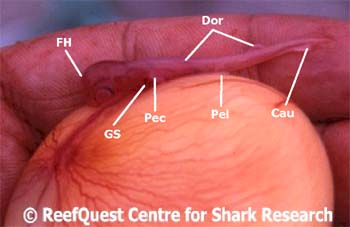 A lateral view of an embryo Green-Eyed Dogfish, outside the
candle. Note the high domed 'forehead' (FH), so unlike the
streamlined shape it would have developed. Note also the tiny gill
slits (GS), the pectoral fin fold (Pec), the pelvic fin fold
(Pel), the dorsal fin fold (Dor), and the developing caudal fin
(Cau), already differentiated into distinct upper and lower lobes.
A lateral view of an embryo Green-Eyed Dogfish, outside the
candle. Note the high domed 'forehead' (FH), so unlike the
streamlined shape it would have developed. Note also the tiny gill
slits (GS), the pectoral fin fold (Pec), the pelvic fin fold
(Pel), the dorsal fin fold (Dor), and the developing caudal fin
(Cau), already differentiated into distinct upper and lower lobes.
