Wound Healing in Sharks
The following reply was given to a list-server inquiry regarding shark wounds and the healing process.
It has long been noted that sharks' wounds heal remarkably quickly. Yet the processes of wound healing in sharks have received surprisingly little scientific attention. This is especially curious, given the intense recent biomedical attention paid the sharks' immune system with regards to cancers (especially tumor angiogenesis) and arthritis as well as metabolic pathways in the liver that detoxify various bacterial, fungal, anthropogenic (man-made, such as estrogen-mimicking organohalides) and environmental toxins (such as bioaccumulation of naturally-occurring lead and other heavy metals).
An important factor in wound-healing in sharks is the fact that their dermal-denticles (like their teeth and fin spines, which are themselves basically highly derived dermal denticles) are continually replaced. Wolf-Ernst Reif, of the University of Tubingen, Germany, is probably the premier investigator of squamation (especially patterns of dermal denticle, fin spine, and tooth growth, differentiation, and replacement) in elasmobranchs. Twenty years ago, Wolf published one of the few studies on wound healing in sharks (Zoomorphologie 90: 101-111). Specifically, Wolf investigated the form and arrangement of repair scales in mature specimens of Nurse (Ginglymostoma cirratum) and Leopard (Triakis semifasciata) Sharks.
Wolf surgically removed 7 by 7-millimetre (about 1/2 inch square) pieces of skin from his test subjects. He noted that during the following two weeks, the wound area secreted mucus, later it contracted and the epidermis (outer skin layer) regenerated; most of the scar area was covered with denticles after 4 months. He also found that the regenerated denticles show a high degree of variability, being much larger and more complex than the scales of control (undamaged) areas. Also, Wolf found that the repair scales were no longer arranged in diagonal rows nor were they oriented in a caudal direction (with the cusps pointing toward the tail), as in the control scales. From these results and a familiarity with Wolpert's (1970, 1972) theoretical work on pattern formation in organisms, Wolf concluded that the flow of positional information was temporarily inhibited by the surgery. (More recent epigenetic [developmental] research has revealed that this positional information is based on concentration gradients of certain molecular markers, the specific markers and their pattern of concentration in turn directed by homeogenes; see Lawrence 1992 for an excellent discussion of this biochemical choreography in the fruit fly Drosophila.)
Wolf's study did not indicate whether the anomalous (weird) replacement denticles were eventually replaced by normal denticles if the flow of positional information is 'readjusted'. In several previously curated skin samples in his collection, however, Wolf found small areas with denticles of normal shapes but oriented anomalously: parallel to one another and forming an angle with the longitudinal (long) axis, so that they no longer pointer toward the caudal (tail) fin. From this observed pattern, Wolf deduced that these angled denticles were traces of healed scars. Wolf concluded that that the size, shape, and variability of the denticles that replace the repair denticles are normal and that the scar area gains a new polarity which is at an angle to the original polarity. But to test fully these conclusions, he noted, a new excision of denticles would have to be made and that it would probably take two years until all the repair denticles were replaced. This work, to the best of my (admittedly incomplete) knowledge, has not been done and would thus make an excellent thesis topic.
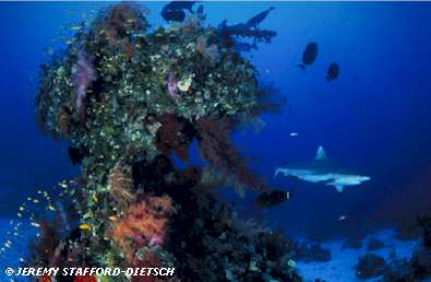
Reef-dwelling sharks, like this Grey Reef Shark (Carcharhinus amblyrhynchos), live in a complex, ever changing environment and despite their phenomenal agility accidents do happen. Most accidents consist of high-speed collisions with reef substrate during pursuit of prey. Fortunately for them, sharks have terrific wound-healing abilities and rarely endure open wounds for long.
Photo © Jeremy Stafford-Deitsch; used with the gracious permission of the photographer, who asks that you support the Shark Trust.
In my own work on the behavioral ecology of reef sharks, I have noted that dermal wounds even ones that appear quite severe often heal amazingly quickly. In 60 to 70-centimetre (roughly two-foot) long juvenile Blackfin Reef Sharks (Carcharhinus melanopterus) observed in estuarine mangrove waters of the central Queensland coast, fresh combat scars (less than two months old) appear whitish, turn black by 4 months, and completely disappear within 6 months. In adults of the Grey Reef Shark (Carcharhinus amblyrhynchos) a 1.2 to 1.5-metre (4 or 5-foot) species observed off Australia, Palau, Fiji, Cook Islands, and French Polynesia individual females with nasty-looking interdorsal wounds (spatiotemporally consistent with courtship scarring) frequently healed in as little as two weeks. Immersion in saltwater or bacteria (notably Clostridium spp., which seem to be normal inhabitants of the mucus coating the mouth, gums, and lips of carcharhinid sharks) introduced into the wounds by courting males or attacking conspecifics may have facilitated and/or accelerated wound-healing in these cases. In subadult and adult Bluntnose Sixgill Sharks (Hexanchus griseus) a 2.5- to 5-metre (8 to 15-foot) coldwater species observed seasonally in relatively shallow waters off British Columbia, Canada white wounds (possibly mating scars or, less likely, combat wounds) of tagged individuals frequently appear, turn dark, and become invisible within a single season (August to November). Klimley and Nelson (1981, 1984) noted similar patterns of wound healing in adolescent female Scalloped Hammerheads (Sphyrna lewini) in the Sea of Cortez. (Such observations on sharks in the wild suggests that temperature may be less important than immersion in seawater in determining the rate of wound healing, but other factors such as feeding success, social rank, bacteria introduced into wounds, etc. may also play a role in determining the rate of wound healing in free-swimming sharks.) I have not yet personally investigated the form and orientation of replacement denticles in any shark species, but plan to begin doing so at my earliest opportunity.
It would be interesting to compare the rate of wound healing in captive versus free-swimming sharks. Sharks are well known to be highly susceptible to the stresses of capture and containment. Pittenger and Gilbert (1986) studied physiological responses to stress in 10 captive adult horn sharks (Heterodontus francisci), held in a 4 000-litre (1 000 gallon) pool. The animals were stressed by removing them from the water and inserting into their caudal vein a catheter back filled with heparinized elasmobranch Ringer's (a physiologically inert saline solution) and extending some 3 metres (10 feet) to float on the surface. Blood samples (0.3 to 0.4 ml) were taken at 15-minute intervals during the first hour after stress and at less frequent intervals thereafter. These blood samples were analyzed for various physiological parameters (lactate, hematocrit, and pH). In all cases, the stressed horn sharks demonstrated rapid rises in lactate (an acidic by-product of anaerobic glycolysis) and hematocrit (the proportion of blood that consists of cells, usually expressed as a percentage) and a lowering of pH (increase in acidity) immediately after stress which persisted for as much as three hours. Captive sharks often exhibit loss of coordination and equilibrium if tank size and/or shape is inadequate for them (captive cruising sharks apparently need long, straight uninterrupted stretches of tank to allow them to alternate between active swimming and passive gliding) and frequently develop Vibrio and other infections and/or persistent wounds on the tip of the snout (from repeated collisions with tank walls). Given the well-documented immunosuppresive effects of stress in other animals, it would be interesting to compare rate of wound healing in stressed versus un-stressed captive sharks, which may allow us to determine the relevant variables and allow us to make testable predictions about wound healing in free-swimming sharks. Perhaps most practically, such research will provide us with the information needed to take better care of captive elasmobranchs.
Cheers,
R. Aidan Martin
Posted to SHARK-L April 8, 1998
